Definition: It is defined as expectoration of the blood from the respiratory tract.
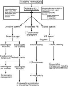
Massive hemoptysis: Massive hemoptysis is broadly defined as production of 500-600 ml of blood over 24 hours; 100 ml within 1 hour or bleeding necessitating transfusion or causing hemodynamic compromise.
Pathogenesis:
- Primary origin of the blood comes from bronchial arteries
- Other sources of bleeding might be:
- aorta
- intercostals arteries
- coronary arteries
- Thoracic arteries
- The upper and inferior phrenic arteries
- Pulmonary vessels
- Hemoptysis may happen following infarction and ischemia of pulmonary parenchyma. It is seen in following conditions:
- Pulmonary emboli
- Vasculitis including:
- Granulomatosis with polyangitis
- Infections including:
- Staphylococcus aureus
- Pseudomonas aeruginosa
- Aspergillus fumigatus
- The phycomycetes
- Another mechanism of hemoptysis is vascular engorgement with erosion. It is seen in following conditions:
- Acute infection such as:
- Viral bronchitis
- Bacterial bronchitis
- Chronic infection such as:
- bronchiectasis
- Toxic exposure
- Cigarette smoke
- asbestos
- Acute infection such as:
- In some cases underlying cause cannot be identified and they are considered as idiopathic. However, they might present with massive hemoptysis.
Causes:
- Bronchial disease:
- Carcinoma
- Bronchiectasis
- Acute bronchitis
- Bronchial adenoma
- Foreign body
- Parenchymal disease:
- Tuberculosis
- Suppurative pneumonia
- Lung abscess
- Parasites( eg: hydatid, flukes)
- Trauma
- Actinomycosis
- Mycetoma
- Lung vascular disease:
- Pulmonary infarction
- Polyarteritis nodusa
- Good pasture syndrome
- Idiopathic pulmonary hemosiderosis
- Cardiovascular disease:
- Acute left ventricular failure
- Mitral stenosis
- Aortic aneurysm
- Blood disorders:
- Leukemia
- Hemophilia
- Anticoagulants
Other history and symptoms accompanying hemoptysis:
- h/o fever- duration, timing and pattern
Suggestive of infective origin (eg: bronchiectasis, lung abscess, tuberculosis, suppurative pneumonia, acute bronchitis)
- h/o cough
- h/o repeated small hemoptysis or blood streaking of sputum
Highly suggestive of bronchogenic carcinoma
- h/o fever, night sweats and weight loss
Highly suggestive of tuberculosis
- h/o rusty colored sputum but may cause frank hemoptysis
Highly suggestive of pneumococcal pneumonia
- Catastrophic bronchial hemorrhage with h/o previous tuberculosis or pneumonia in early life
Highly suggestive of bronchogenic carcinoma
- h/o prolonged immobilization, malignant disease of any organ, cardiac failure and pregnancy
Highly suggestive of pulmonary thrombo embolism
On examination:
- Anemic
- Temperature (usually high)
- Clubbing of fingers (highly suggestive of bronchogenic carcinoma or bronchiectasis)
- Cachexia, hepatomegaly with lymphadenopathy: highly suggestive of bronchogenic carcinoma
- Fever, signs of consolidation and pleurisy: highly suggestive of pneumonia, pulmonary infarction
- Signs of cavitation, consolidation and pleural effusion: highly suggestive of tuberculosis
- Legs signs of deep vein thrombosis: highly suggestive of pulmonary infarction
- Rashes, hematuria, splinter hemorrhages, lymphadenopathy or spleenomegaly: highly suggestive of systemic diseases
Investigations:
- Complete blood counts with ESR
- Chest x- ray (gives clear evidence of localized lesion including pulmonary infarction, tumor- malignant or benign, pneumonia, tuberculosis
- Sputum for AFB, gram stain and culture
- Mantoux test
- CT scan of chest and CT guided FNAC from a mass lesion
- Hematological test including bleeding and clotting time
- Bronchoscopy for bronchogenic carcinoma
- Ventilation perfusion lung scan for pulmonary thromboembolic disease
Treatment:
- If massive hemoptysis :

- Otherwise treatment should be according to the cause
Difference between hemoptysis and hematemesis
| HEMOPTYSIS | HEMATEMESIS |
| Usually preceeded by coughing | 1)usually preceeded by vomiting |
| Color: bright red | 2)color altered (dark red or coffee ground) |
| Blood may be associated with frothy sputum | 3)blood may be mixed with gastric content |
| Reaction: alkaline | 4)reaction: acidic |
| h/o respiratory or cardiovascular disease | 5)h/o gastrointestinal disease |
| confirmation with bronchoscopy | 6)confirmation by endoscopy |
Contributor- Dr. Bidhata Rayamajhi


Nicely written 👍👍
👌